Main Second Level Navigation
Breadcrumbs
- Home
- Research
- Facilities and Equipment
- Imaging
Imaging
DCM hosts cutting edge instrumentation such as the Bruker Xtreme and IVIS Spectrum to meet your imaging needs.

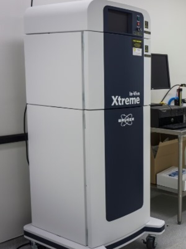
Bruker Xtreme
The Bruker Xtreme instrument provides in-vivo optical and x-ray imaging including four modalities: high resolution X-ray, fluorescence, luminescence and radioisotopic. By overlaying high sensitivity optical imaging with high resolution X-ray, you can achieve precise anatomical localization of your molecular or cellular biomarker. In addition, the unit offers DEXA scanning and bone density analysis. A built in anesthetic system supports high throughput imaging of up to 5 mice at the same time with dedicated mouse housing available nearby for serial imaging over the duration of your study.
The multimodal imaging system can be configured to support optical imaging of bacterial and viral markers, molecular indicators of apoptosis or inflammatory processes in detailed co-registered anatomical context, multiple animal organs or tissues can also be imaged simultaneously.
A dedicated Class II Type A2 supports work with RG2 agents. Use the Xtreme to image real-time changes in biochemical pathways, quantify changes in bone and soft tissue structure or track migration of cells longitudinally.
CCD Camera
- Back-thinned, back-illuminated 4MP CCD detector
- 13.5 micron pixels, 2048 x 2048
- Large, fast f/1.1 – f/16, 58 mm lens, fixed
Luminescence Sensitivity
- BI 4MP: <650 photons/sec/cm2 and <50 photons/sec/cm2/
Fluorescence Light Source & Excitation Filters
- Powerful 400W Xenon illuminator
- 28 narrow band excitation filters
- Excite fluorophores from the visible to the NIR, 410-760nm
- 6 high-sensitivity wide angle emission filters, automated 8 position filter wheel, 535-830nm
X-ray Source
- Geometric magnification stage for high resolution imaging
- High speed X-ray head 500 μA
- 20-45kVp
The Xtreme is professionally managed through the 3D-CFI facility offering full image analysis services as well as training should you wish to do the imaging yourself.

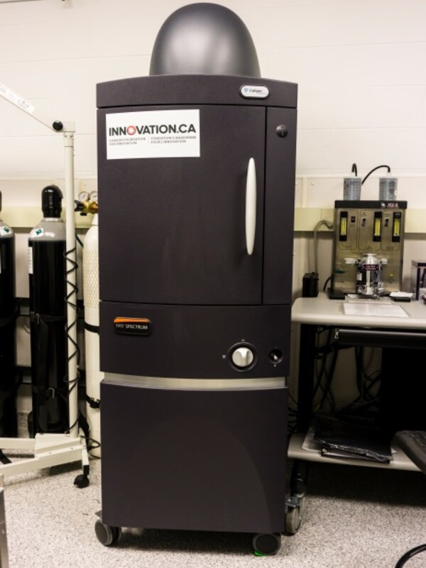
IVIS Spectrum
The IVIS Spectrum instrument provides high sensitivity in-vivo optical imaging via three modalities: fluorescence, luminescence. The Spectrum also offers full 3-D tomographic anatomical reconstruction of your fluorescent or bioluminescent reporter against the built-in Digital Mouse Atlas. An integrated anesthetic system supports high throughput imaging of up to 5 mice at the same time with dedicated mouse housing available nearby for serial imaging over the duration of your study.
Image multiple fluorescent probes and bioluminescent reporters like firefly luciferase, Renilla luciferase and bacterial luciferase in vivo at depth rapidly and quantitatively. A dedicated Class II Type A2 supports work with RG2 agents. The ultra-sensitive camera allows for detection of as few as five cells for applications such as early monitoring of metastasis, longitudinal tracking of tumor development or stem cell tracking in whole animal body.
Image and quantify all commonly used fluorophores, including fluorescent proteins, dyes and conjugates. Automatic Spectral unmixing allows detection and separation of multiple reporters, and reduces the effects of tissue auto-fluorescence, permitting visualization of reporters in various applications such as tracking cell recruitment in inflammation.
CCD Camera
- Back thinned, back illuminated grade 1 CCD provides high quantum efficiency over the entire visible to near-infrared spectrum
- 16-bit digitizer delivers broad dynamic range
- 13.5 micron pixels, 2048 x 2048
- CCD is thermoelectrically (Peltier) cooled to -90 °C , ensuring low dark current and low noise
Custom-Designed Lens
- • 6-inch diameter optics, f/1– f/8
- • High-resolution - down to 20 microns
- f/1 - f/8 lens; 1.5 x, 2.5 x, 5 x, 8.7 x magnifications
Fluorescence Light Source and Excitation Filters
- Emitted light from excitation filter wheel
- 10 narrow band excitation filters: 415 nm -760 nm
- 18 narrow band emission filters: 490 nm -850 nm
The Spectrum is professionally managed through the 3D-CFI facility offering full image analysis services as well as training should you wish to do the imaging yourself.

X-ray
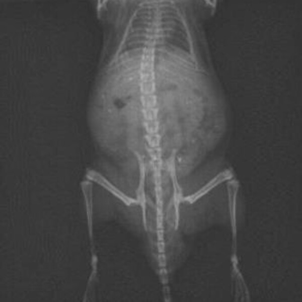
The Min X-ray SA100 portable unit offers high resolution x-ray imaging with AGFA CR30 Xe digital MUSICA™ processing and image analysis with NX software. Zoom in and adjust image contrast, frequency and density for optimal feature identification and viewing on the 2MP monitor. Images in DICOM or jpeg format burned to disk for your files. Track the progress of your orthopedic project from immediate post-operative imaging through critical periods of change.
